

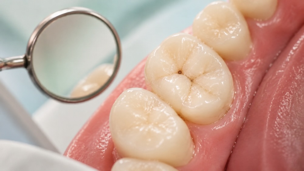
A cavity may first look like a chalky white or dull spot where enamel is losing minerals. As decay progresses, the area can become yellow, brown, gray, or black and may develop a rough pit, indentation, or visible hole. Some cavities, especially those between teeth or beneath existing dental work, cannot be seen without a dental exam and X-rays.
A photo can help you recognize a change worth checking, but it cannot confirm tooth decay. Stains, tartar, naturally dark grooves, and enamel defects can resemble a cavity. If you notice a new spot, rough area, food trap, or sensitivity, a dental exam is the reliable next step.
What Does a Cavity Look Like? The Short Answer
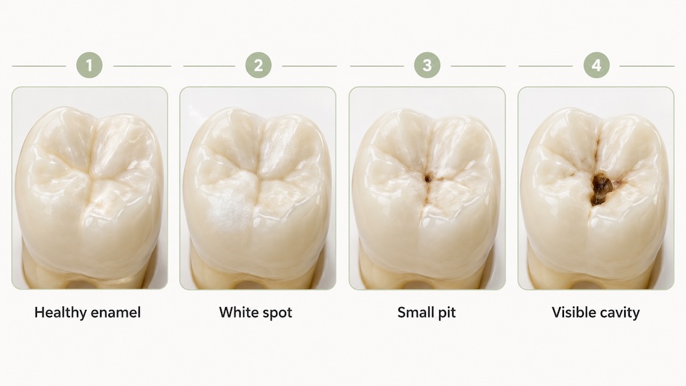
Depending on its stage and location, tooth decay may look like:
- A matte, opaque, or chalky white patch
- A localized yellow, brown, gray, or black spot
- A rough area that catches food or floss
- A small pit or indentation
- A visible hole with missing tooth structure
That list is useful for comparison, not diagnosis. A black mark does not automatically mean severe decay, and an early cavity may have no dark color or visible hole at all.
What Cavities Look Like at Each Stage
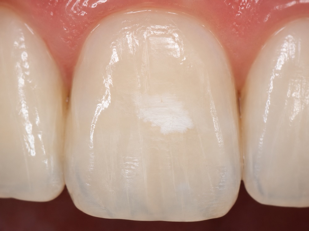
Stage 1: Early demineralization or a white spot
When a cavity first starts, the enamel may look dull, matte, or chalky instead of smooth and glossy. The affected area is often white because minerals have been lost from the enamel. There is not yet a physical hole.
This distinction matters. The National Institute of Dental and Craniofacial Research explains that early mineral loss may be stopped or reversed before the enamel surface collapses. A dentist may recommend fluoride, better plaque control, diet changes, and monitoring based on your individual risk. Our preventive dentistry services are designed to identify and manage problems at this earlier stage.
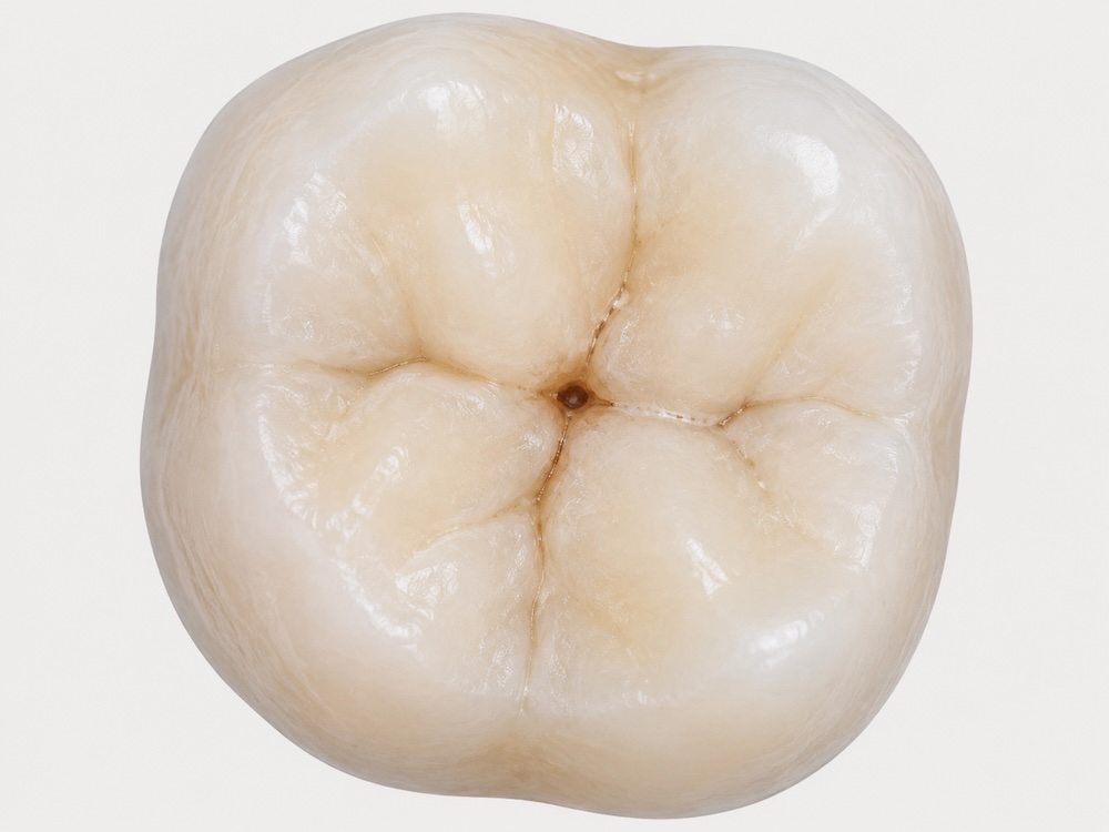
Stage 2: Enamel breakdown and a small pit
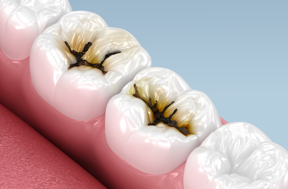
As enamel weakens, a small rough spot, pit, or indentation may develop. On a molar, it can appear as a tiny brown or black point in one of the natural grooves on the chewing surface. However, many healthy grooves are naturally dark or stained. Color alone cannot tell you whether the enamel is actively breaking down.
Stage 3: Decay reaches the dentin
Dentin is the layer beneath the enamel. It is softer than enamel, so decay can spread more quickly after reaching it. The area may look wider, darker, or more hollow, with visible loss of tooth structure. You may also begin to notice sensitivity to cold, sweets, or pressure, although some cavities still cause no symptoms.
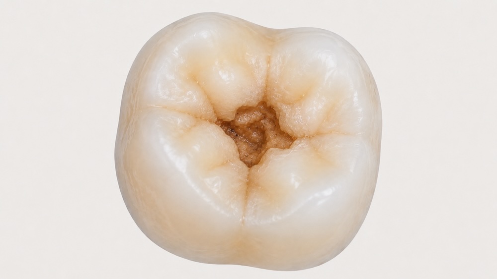
Stage 4: Deep decay approaches the pulp
A deep cavity may look like a large brown or black opening, a broken edge, or a section of missing tooth. When decay approaches the pulp, where the tooth’s nerves and blood vessels are located, symptoms can include lingering sensitivity, spontaneous pain, pain at night, or pain when biting.
Deep decay does not automatically mean the tooth needs to be removed. Depending on the amount of healthy structure remaining and the condition of the pulp, treatment may involve a larger restoration, a crown, or root canal treatment.
Stage 5: Infection or abscess
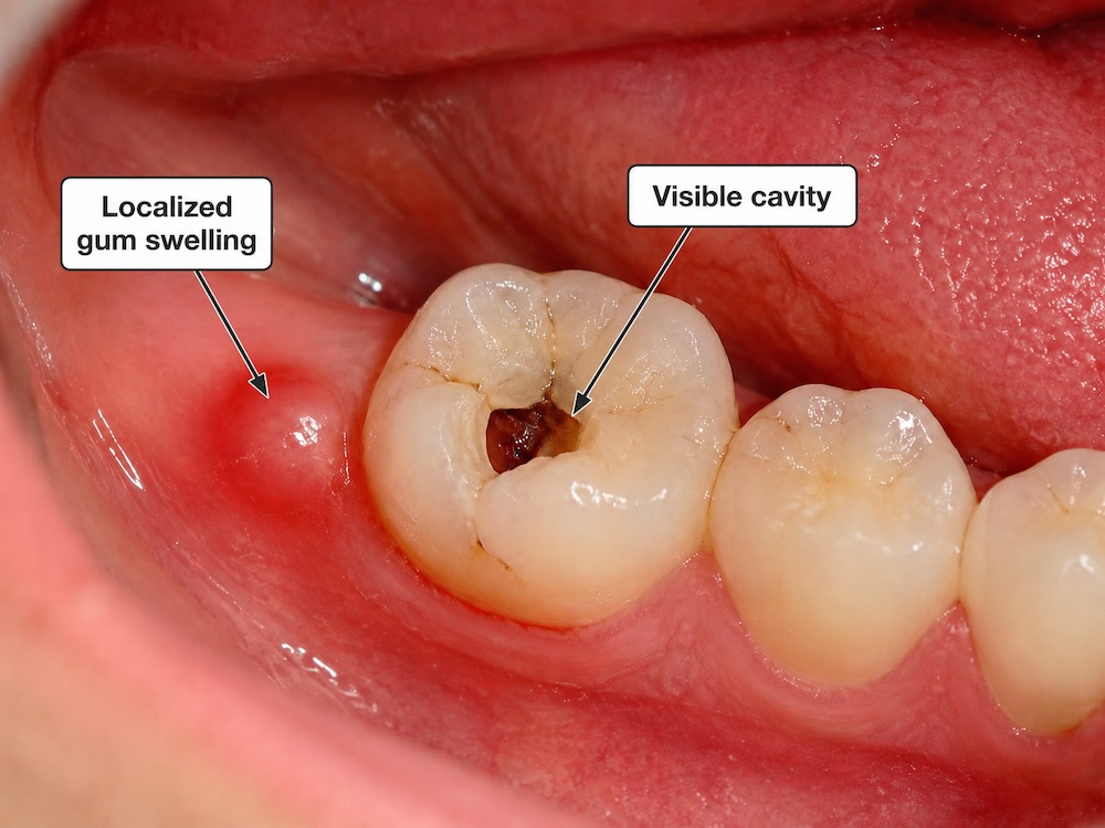
An abscess is an infection, not simply a darker-looking cavity. Warning signs can include a pimple-like bump on the gum, drainage, a bad taste, throbbing pain, fever, or swelling in the gums, jaw, or face. Some infected teeth may hurt less if the nerve has died, so reduced pain does not necessarily mean the problem has resolved.
Get prompt dental care for swelling, fever, drainage, severe or spreading pain, or difficulty opening your mouth. Trouble swallowing or breathing and rapidly increasing facial swelling require emergency medical attention. Green California Dental offers emergency dental care in the Los Angeles area for urgent dental problems.
What Cavities Look Like in Different Places
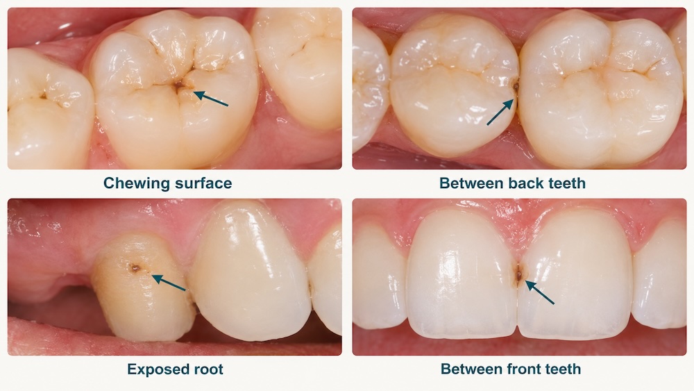
On a molar’s chewing surface
Molar cavities often begin in pits and fissures, the narrow grooves that collect plaque and food. You might see a tiny dark point, a rough opening, or a larger area where enamel has broken away. A dark groove may also be harmless staining, which is why a dentist evaluates the surface rather than judging it only by color.
Between teeth
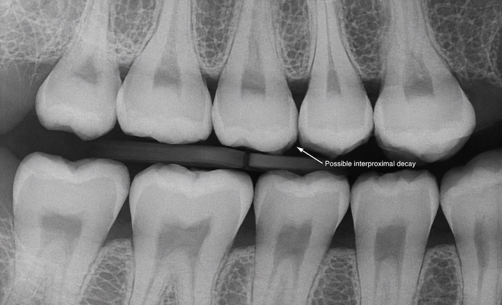
A cavity between teeth often cannot be seen in a mirror. It may first become noticeable because floss catches or tears in one area, food repeatedly packs between two teeth, or a tooth becomes sensitive. On a bitewing X-ray, decay may appear as a darker area where the tooth structure has lost density.
Near the gumline or on an exposed root
Root decay may appear yellow-brown, tan, or dark near the gumline, especially where gum recession has exposed the root. Root surfaces are not covered by enamel, so decay can progress differently and may need attention sooner.
On a front tooth
On front teeth, early decay may appear as a white spot near the gumline. Decay between front teeth can look like a localized gray or brown shadow. A chip, developmental enamel defect, or surface stain can look similar, so do not use appearance alone to choose a treatment.
Cavity or Stain? Why Color Alone Is Not Enough
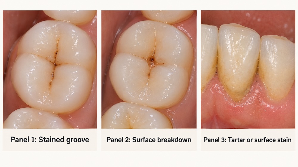
A stain changes the color of the tooth surface, while active decay involves mineral loss and, in later stages, breakdown of tooth structure. The problem is that they can look similar at home.
- Surface stains from coffee, tea, tobacco, or highly pigmented foods may affect several teeth or follow broader surface patterns.
- Stained molar grooves can look like thin brown or black lines without active decay.
- Tartar can appear yellow, brown, or dark near the gumline and cannot be brushed away.
- Fluorosis or enamel hypoplasia can create white, cream, yellow, or brown areas that are not cavities.
- Old fillings and sealants can change color or create visible margins that need professional evaluation.
A dentist considers the surface texture, location, changes over time, symptoms, cavity risk, and imaging when appropriate. There is no reliable at-home color test.
Can You Have a Cavity You Cannot See?
Yes. Decay can develop between teeth, beneath an old filling or crown, or on the back side of a tooth where a mirror and phone camera cannot reach. Even a dentist cannot see every area directly during a visual exam.
Dental X-rays can help identify disease that is not visible during a regular examination, according to the American Dental Association. X-rays are not taken on the same schedule for everyone. A dentist determines when they are appropriate based on age, current oral health, cavity risk, symptoms, and previous findings.
What a Cavity May Feel Like
Early decay commonly causes no symptoms. As it progresses, you may notice:
- Sensitivity to sweets, cold, or heat
- Pain when biting or chewing
- Food catching in the same spot
- A rough edge or hole you can feel with your tongue
- Persistent or throbbing tooth pain
- A bad taste, drainage, or swelling
These symptoms are not specific to cavities. Cracks, worn enamel, gum recession, a loose filling, and other dental problems can feel similar. You can also read our article on what a cavity can feel like to gather more information.
When to Schedule a Dental Exam
Schedule an exam if you notice a new white, brown, gray, or black area; a pit or rough spot; repeated food trapping; a broken filling; or new sensitivity. Waiting for pain is a poor test because enamel decay may be painless, and some advanced problems do not produce constant symptoms.
If you have severe pain or swelling, contact a dentist promptly. For a practical overview of other situations that should not wait, read our guide to the top reasons to visit an emergency dentist immediately.
How Dentists Confirm and Treat a Cavity
A dentist confirms tooth decay by combining your history and symptoms with a clinical examination, cavity-risk assessment, and X-rays when indicated. The goal is to determine whether the area is active, whether the enamel surface is still intact, and how deeply the problem extends.
Treatment depends on the stage:
- Early mineral loss without a hole: fluoride, improved home care, dietary changes, and monitoring may help arrest or reverse the process.
- A cavitated tooth: damaged structure usually needs to be removed and repaired with a filling or another restoration. Learn more about our direct restorative dentistry options.
- Extensive structural damage: the tooth may need an inlay, onlay, or crown to restore strength and function.
- Decay involving the pulp: root canal treatment may be needed to remove infected or inflamed tissue and preserve the tooth.
Repairing decay can also improve the look and function of a damaged tooth. Our related article explains how dentistry can address tooth damage, loss, or decay.
How to Lower Your Risk of New Cavities
- Brush twice a day with fluoride toothpaste.
- Clean between your teeth every day with floss or another tool recommended by your dental team.
- Limit how often you sip or snack on sugary and starchy foods and drinks.
- Ask about fluoride treatments or sealants if you are at higher risk.
- Address dry mouth, which reduces saliva’s protective effect.
- Keep preventive visits on the schedule your dentist recommends for your risk level.
Frequently Asked Questions
How can I tell if it is a cavity or not?
You cannot confirm a cavity from appearance alone. A localized white, brown, or black change, a rough spot, sensitivity, or food trapping can justify an exam, but stains and other enamel changes may look similar. A dentist may use an examination and X-rays when indicated.
What does a stage 1 cavity look like?
The earliest stage often looks like a matte, opaque, or chalky white spot caused by mineral loss. The enamel surface is still intact, so there is no visible hole. This stage may be arrested or reversed with professional guidance.
What does a small cavity look like?
A small cavity may look like a tiny pit, rough area, or localized brown or black point, especially in a molar groove. It may also be invisible if it is between teeth.
Are all black spots on teeth cavities?
No. A black spot may be a stain, tartar, an old filling, or a naturally dark groove. Some dark areas can represent decay, but a dentist must examine the tooth to tell the difference.
What does a cavity look like on an X-ray?
Tooth decay often appears as a darker, or radiolucent, area where tooth structure is less dense. Between teeth, it may form a small triangular shadow. X-rays must be interpreted with the clinical exam.
Can a cavity heal on its own?
Early mineral loss may be stopped or reversed before a hole forms. Once the tooth surface has collapsed into a true cavity, the missing structure does not grow back and usually requires a restoration.
How late is too late for a cavity?
There is no safe visual cutoff. Earlier care is usually simpler. Pain, drainage, fever, or swelling needs prompt evaluation, and difficulty swallowing or breathing requires emergency medical attention.
Book a Cavity Check with Green California Dental
If you are unsure whether a spot is a cavity, schedule an exam with Green California Dental in Burbank. We can inspect the tooth, take X-rays when appropriate, and explain the least invasive next step based on what we find.